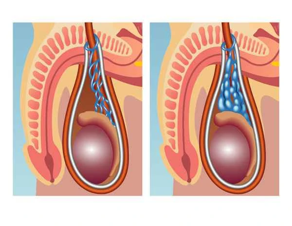
An ultrasound examination for varicocele is a medical examination that uses ultrasound technology to observe the condition of the spermatic veins in the male reproductive system. This examination is crucial for diagnosing varicocele because it helps doctors accurately determine whether the spermatic veins are dilated and to what extent, thus providing patients with more effective treatment options.

During an ultrasound examination for varicocele, doctors use ultrasound imaging technology to observe the structure and blood flow of the spermatic veins. This examination not only visually displays the morphology of the spermatic veins but also assesses the degree of venous dilation by measuring the vein's diameter. By observing the direction and speed of blood flow, doctors can further determine if there are problems such as blood reflux, which is crucial for developing a treatment plan.

Examination results are usually presented in numerical form, such as the specific value of the vein diameter. Generally, a vein diameter exceeding the normal range may indicate the presence of varicocele. It is important to recognize that these numbers are only one part of the diagnosis and need to be considered in conjunction with clinical symptoms and other examination results. When faced with examination results, patients should maintain an objective attitude, avoiding over-interpreting the meaning of the numbers themselves, and instead focusing more on the doctor's overall assessment and recommendations.
【Useful Tips】
1. Before undergoing an ultrasound examination for varicocele, try to remain calm and avoid strenuous exercise.
2. During the examination, follow the doctor's instructions and maintain an appropriate posture to obtain clear images.
3. After receiving the results, communicate fully with the doctor to understand the specific meaning of the test results and subsequent treatment suggestions.
4. Follow your doctor's instructions, have regular check-ups, and adjust the treatment plan as needed.